

© 2024 AT Home Care & Hospice
All Rights Reserved
All Rights Reserved

By: By Babatope Olusina, PT, DPT and Olaide Oluwole-Sangoseni, PhD, DPT, MSc.
Osteoarthritis is a degenerative joint disorder that affects the articular cartilage, underlying bone, and surrounding soft tissues. It is the most common form of joint disease in the United States (US), with an estimated prevalence of 27 million people,1 with an occurrence of about 10% in men and 13% in women, over the age of 60 years. 2 Hip osteoarthritis accounts for about 70% of total hip arthroplasties (THA) that are performed in the US due to severe pain, which limits the individual’s functional mobility and negatively affects his/her activities of daily living (ADLs), eventually limiting his/her participation in work and leisure activities. 3 Other indications for THA include but are not limited to trauma and osteonecrosis of the femoral head.4
THA is the surgical replacement of the natural hip joint with a prosthesis. 3 The first THA procedure was completed in the US in 1969 and as the procedure has grown in incidence, the technique has evolved and its efficacy has improved. 2,4 A 2010 prevalence study estimated 2.34% of individuals over the age of SO years in the United States have had THA, corresponding to 2.5 million people (1.4 million women). 4 A detailed breakdown of their study revealed a prevalence of 0.58% at age SO years, increasing to 1.49% at sixty years, 3.25% at seventy years, 5.26% at eighty years, and 5.87% at ninety years of age.4 The original or more popular technique is the posterior or posterolateral approach, with its associated precautions (no hip flexion above 90 degrees, no adduction beyond the midline, and no internal rotation of the surgical hip joint).
In the 1980s, an anterior approach was developed and gained popularity because of improved early outcomes in terms of pain and early functional recovery. 5 Hip hemiarthroplasty is the surgical removal of one of the components of the hip joint, most often the femoral head. Although it is less invasive, the Physical Therapy assessment and management will follow a similar path as for a THA.
Home health physical therapists (HHPT) are part of the multidisciplinary team-approach called upon to manage these patients upon their return home. Orthopedic surgeons seek the involvement of physical therapists (PT), as movement specialists, to facilitate the recovery and rehabilitation of THA patients to maximize their return to full function and participation in the activity. In consultation with the orthopedic surgeons, our home health agency established protocols that can be customized to fit the individual patient’s desired outcomes and surgeon’s preferences. These protocols guide the first few weeks of in-home rehabilitation before the transition to outpatient physical therapy. As a HHPT with a weekly caseload of about 50 percent of total hip and total knee arthroplasty patients, I recognize that no two patients’ status post-THA is the same.
The Medicare home health Conditions of Participation (CoP) require that a comprehensive assessment of each patient be performed by the admitting clinician to start the episode of care. This assessment includes the patient’s past medical history (PMH); a complete review of the patient’s medications, including any changes in dosage and patient’s response; and the integumentary assessment, all of which go into the process of formulating the patient’s plan of care.
Because most home health patients have multiple diagnoses, the evaluating PT incorporates the patient’s past medical/surgical history and the patient’s prior level of function into his/her physical therapy plan of interventions. A detailed assessment of the patient’s home is an essential aspect of the initial visit, as safety hazard/fall risks can be identified, and safety education and recommendations can be immediately communicated to the patient and their caregivers. The purpose of this case report is to highlight the physical therapy management of a THA in the home health setting with a focus on adapting exercise program based on pain and muscle fitness indices.
The patient is an 81-year-old female, retired nurse admitted to HHPT following right THA revision with weight-bearing as tolerated precautions. She resides alone in a single level house in a 55+ Age-in-Place community. Before this surgery, she was fully independent with all of her functional mobility, occasionally using a standard straight cane for ambulation. She was independent with all activities of daily living (ADL)s and was active in her community, participating in group exercise sessions every week.
Her PMH was significant for multiple comorbidities: hypertension, atrial fibrillation, type 2 diabetes mellitus, anxiety, neuropathy, general osteoarthritis, vertigo, history of lumbar laminectomy and fusion in 2017, reflux disorder, cystocele with prolapse, and recent urinary tract infection. Initial THA was performed in 2002; she had a dislocation in 2017, which resulted in persistent hip joint pain afterward.
• Apixaban, PO, 2.5mg, 1 tab, twice daily
• Diltiazem, PO, 240mg, 1 tab daily
• Norvasc, PO, 5mg, 1 tab PRN, if diastolic is above 90mmHg
• Pepcid, PO, 10mg, 1 tab Q 8 hours
• Xanax, PO, 0.5mg, 1 tab daily
• Tramadol, PO, 50mg, 1 tab Q 6 hours
• Purelax PO, 17gram/dose, 1 tab twice daily
• Potassium Chloride, PO, 20mEq, 1 tab twice daily.
• Tylenol Extra strength, PO, 500mg, 2 tabs Q 8 hours
The patient stated her goal is to return to walking independently without the walker.
On examination, “Nanette”(a pseudonym) was alert and oriented to person, place, and time, and able to follow a multi-level command. She presented with hypomobility of the hip joint and weakness of the proximal muscles of the surgical lower extremity. She required contact guard/minimal assistance to assist her right lower extremity into the bed during bed mobility assessment. She required close stand-by-assistance of the therapist for sit to/from stand and bed to/from chair transfers due to She was dependent on a rolling walker for ambulation, with forward-flexed trunk posture over the device, using a 2-point antalgic gait pattern. The Timed Up and Go test (TUG) was performed, with the “Nanette” requiring 36 seconds to complete. 6 A time greater than 20 seconds is indicative that the patient is dependent on transfers and confirms homebound status. 6 The family had hired a private duty company to provide personal care assistance for several hours during the day to assist her in the first 3 weeks.
Because the initial home PT evaluation was on the same day as the nursing start of care (SOC) visit, PT evaluation referred to the nursing integumentary assessment. However, the physical therapist assessed the surgical incision on each subsequent visit, per our agency protocol, performing dressing changes when needed. The status of the wound was documented and presented at the interdisciplinary case conference with the registered nurse as required.
Per Medicare CoP, Nanette’s pain was evaluated on the initial examination and all subsequent visits, using a numeric pain rating scale (0-10) and verbal description. She rated her pain at level 5/10; her goal was to be pain-free by discharge. The expectation was a gradual decrease in the pain level, and any movement in the other direction and/or a new type of pain warrants additional evaluation.
Based on the hypomobility of the hip joint and weakness of the proximal muscles of the hip and pelvis, the International Classification of Functioning, Disability and Health (ICF) model diagnosis of M25.651 or 652 (stiffness of hip joint, not elsewhere classified) was made.
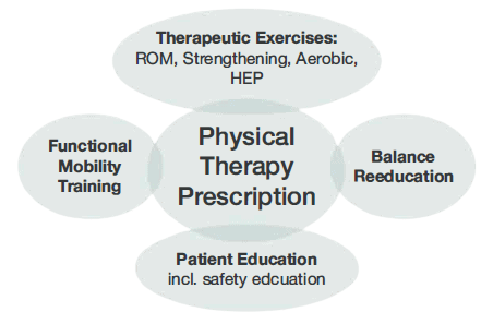
It was determined that Nanette would benefit from a skilled PT intervention frequency of 2 times per week for 4 weeks. Her exercise prescription consisted of joint mobilization, ROM and strengthening exercises, instruction in a home exercise program (HEP), functional mobility training (including bed mobility, transfer, and gait training), balance reeducation, equipment training, safety education, to progress to independence in all areas (Figure 1).
Since the pain was the chief complaint, it was used as one of the outcome measures. Pain medication was to be taken an hour before the PT session. The patient’s pain level was expected to decrease to level 1/10 at discharge.
The patient achieved independence with her bed mobility by the first session of week 2 (3rd visit), and independence with her sit to/from stand and chair to/from bed transfers by the end of the second week (4th visit). She demonstrated independence with her shower stall and tub transfers by the third week (6th visit) and independence with car transfer in the 4th week. The patient progressed to gait training with a standard cane by the 3rd week. She had progressed to independence with gait training on level and uneven surfaces, including on her inclined driveway and to her community mailbox, which is about 450 feet away from her front door, with a numeric pain score of 0/10. Her functional score improved from 36 seconds at initial evaluation to 12 seconds using the TUG test. This score correlates with independence in all transfers and activities of daily living. Her right hip muscle strength had improved to 3+/5, and the patient demonstrated independence with her HEP. Nanette had achieved pain-free status by the final visit.
This patient presented with several impairments that created limitations at the body function level as well as at the activity and participation level, 11,12 as laid out above with the ICF model.
The HHPT used his/her expertise to provide an individualized, person-centered, evidence-informed treatment in the management of this patient. The plan of care was based on the interdisciplinary protocol developed by the surgeon and the home health agency, to help achieve the goals she stated as important to her. Nanette was motivated to return to driving and get back to her normal social life with her friends in the 55+ Age-in-Place community where she resides. The patient had multiple episodes of elevated blood pressure that warranted the notification of her primary care physician (PCP). To err on the side of caution, our home health agency policy requires physician notification for any systolic above 150 and diastolic above 90mmHg. Although her PCP decided not to take any immediate action, her blood pressure was carefully assessed at the start of each visit and response monitored throughout the interaction. The patient had a medical history of anxiety, which sometimes played a role in her exercise response and feedback to the therapist, especially related to her RPE rating and response. She required frequent reassurance, and it was well managed afterward.
Further, consistent with her medical history, constant adjustments had to be made based on observations and findings at the beginning of each visit. Some of her exercises were modified, considering her history of back pain and surgery to avoid aggravating old symptoms. She received a reiteration of education regarding home safety and community re-entry in the last week of the HHPT visit.
Nanette was discharged from home health nursing services in her second week and was discharged to outpatient therapy upon completion of her 8th and final visit. She planned to start driving, first inside her community, once she was released from home health services. An anonymous satisfaction survey was mailed to her by the agency after discharge, and she expressed her satisfaction to the therapist on her last visit, showing how quickly she was progressed to independence with physical therapy. This case report demonstrates a successful progression of an individual with a posterior approach THA from acute care to return to community living through a three-week course of HH services.
About the Authors

Babatope Olusina, PT, DPT, Certificate of Advanced Competency in Home Health (APTA Home Health Section). He is a member of the APTA and Home Health section. Dr. Olusina now works for a home health agency in Richmond, VA, but previously owned and ran a Contract Therapy Staffing Company.

Dr. Olaide Oluwole-Sangoseni, PhD, DPT, MSc, GCS, is an associate professor of physical at Maryville University of St. Louis. She is a Board Certified Geriatric Specialist and a home health PT. Dr. Sangoseni is an advanced physical therapy clinical specialist degree in neuro-orthopedics from the University College London, England. She is an APTA credentialed clinical instructor. She can be reached at osangoseni@maryville.edu.
References
By: By Babatope Olusina, PT, DPT and Olaide Oluwole-Sangoseni, PhD, DPT, MSc.
Osteoarthritis (OA) is a degenerative joint disorder that affects the articulating bones, the articular cartilage, and surrounding soft tissues. Other causes of joint pain and degeneration include rheumatoid arthritis (RA), osteonecrosis, post-traumatic degenerative joint disease, and other pathologic conditions. While joint pain is usually the first sign that prompts the decision to seek medical attention1, other symptoms soon develop, including but not limited to joint hypomobility and muscle weakness. The progression of the disease results in the development of functional limitations such as difficulties with transfers, gait abnormality, stair management, and completing activities of daily living (ADLs), eventually having negative impacts on work, pleasure, and quality of life.
The knee joint is the largest weight-bearing joint in the body, and chronic knee pain has been listed as a “leading cause of musculoskeletal disability in the United States (US)”. 2 Total knee arthroplasty (TKA) is the surgical replacement of the natural knee joint with a prosthesis. It was the first performed in the US in 1968, and with ongoing advances in the technique and materials used, significant progress has been made in its effectiveness and success rate. A 2010 prevalence study of TKA by Kremers el al3 estimated 1.52% in the entire US population, and 4.55% in those over the age of 50 years, have had TKA. Prevalence is higher in women as compared with men, and it increases with age. Inacio et al4 projects that the number of TKAs performed in the US will increase by the year 2050 to a prevalence of 2.58%, amounting to 2,854 procedures per 100,000 US citizens. Partial knee replacement is often performed when the degenerative changes are confined to a particular compartment of the knee and mostly in the younger patient. The PT management is similar to that of a TKA, as described later in this study.
Physical therapy (PT) plays a major role in the initial conservative management of the knee pain before TKA, not only for pain management but also to improve function and decrease the limitations afflicted by the condition. Following a TKA, the home health physical therapist is part of the multidisciplinary team approach that manages the patient upon discharge home. Our Home Health Agency (HHA) has developed protocols to manage these patient populations effectively. Based on previously established protocol with the referring Orthopedic Surgeon, a registered nurse (RN) completed the initial visit, performing a comprehensive assessment of the patient, including the past medical history (PMH), assessment and care of the surgical incision, medication review/training, and the OASIS elements. This HHA also has established protocol with other Orthopedic Surgeons where the PT is the admitting clinician. The initial PT evaluation consists of a detailed musculoskeletal assessment of the patient,
a review of the patient’s PMH, gait and balance evaluation, and a home safety evaluation. A PT plan of intervention is formulated to address noted impairments and functional limitations, with the patient’s self-stated goal as the endpoint.
The purpose of this case report is to highlight the physical therapy management of TKA in the home health setting.
The patient is a 50-year-old female office worker referred to home PT and Nursing following a right TKA revision due to instability and eventual failure of hardware. She resides with her husband in a single-level house, with 8 entrance steps, and she was fully independent with all of her functional mobility, including ambulating without an assistive device, but she was limited by right knee pain and the knee “locking up”.
Her PMH was significant for multiple comorbidities: Significant for Hypertension, Diabetes Mellitus, Hyperlipidemia, Asthma, Anemia, Cervical spine stenosis, Elevated Hemoglobin, Ehlers-Danlos syndrome, Depression, Metabolic syndrome, and Premature Ventricular Contraction (PVC). Her past surgical history is significant for Bilateral TKA, Anterior cervical discectomy with fusion, and Caesarean Section.
During her initial evaluation completed on 12/26/20, Valerie presented an alert and oriented to person, time, and place, and able to follow multi-level commands. She presented with hypomobility of her right knee (flexion ROM of 81 degrees and extension at negative 6 degrees) and weakness in her right quadriceps and hamstring muscle strength (2+/5 on the Manual Muscle Testing grade). She had difficulty with her bed mobility requiring close stand-by assistance, and she required supervision for her transfers. She was dependent on a pair of axillary crutches for ambulation using a 2-point gait pattern. She also used an antalgic gait pattern and exhibited poor arthrokinematics in the right knee. Her balance was assessed with the Timed Up and Go test (TUGT)5, with a score of 17 seconds; this identified her as having a high risk of falls.
The patient’s right knee surgical incision was covered with “Aquacell”, a non-removable dressing on the day of PT evaluation. It was removed by skilled nursing during the subsequent visit on 12/29/20. PT assessed the patient’s surgical incision during all follow-up visits, for signs and symptoms of infection. This is part of agency protocol that all clinicians will assess surgical incision and document appropriately; coordination of care is performed weekly with the RN case manager.
The patient’s pain level and description were assessed and documented during the initial PT evaluation and subsequent visits. The PT provided education to the patient and her husband regarding pain management strategies with her prescription analgesics, cryotherapy, and movement.
Based on the hypomobility of her Right knee joint and the weakness of her right hamstring and quadriceps muscles, the International Classification for Functioning, Disability and Health (ICF) model ICD diagnosis of M25.661 (stiffness of right knee, not elsewhere classified), was made.
PT determined that patient will benefit from skilled intervention with a frequency of 3 times per week for 3 weeks, per previously established protocol with referring surgeon. The intervention included therapeutic exercises (including a range of motion [ROM], strengthening exercises, and joint mobilization), functional mobility training (including bed mobility, transfer, gait, and stair training), instruction in-home exercise program, balance reeducation, safety education, equipment training, patient and caregiver education, and training, to progress her to independence in all areas.
Days 1-10 acute phase (with emphasis on ROM, isometric and isotonic exercises)
Days 11-21 the sub-acute phase (with progression to advancing ROM, strengthening exercises, and joint mobilization)
Home exercise program (HEP) – the patient was instructed to perform her HEP 2-3 times daily. HEP was upgraded from Phase 1 to Phase 2 as the joint effusion and pain decreased, the right LE muscle strength and coordination improved.
The patient completed 8 of 9 planned HHPT visits, and she had progressed to independent bed mobility and transfers by her third visit on 12/30/21. By the end of the 3rd week her progress was as follows:
The patient’s plan of care was developed based on a combination of her impairments following her right TKA, functional limitations, and goals. She made steady progress towards her stated goals of independence with all of her functional mobility and all of her ADLs, without pain and locking up of her Right knee joint. The patient regained full AROM of her right knee joint and regained muscle strength to engage in her regular activities. She progressed to and demonstrated independence with all of her mobility and planned to resume telecommuting the following week once cleared by her surgeon.
During HH services, the patient was diagnosed with postoperative anemia by her PCP, which is common among surgical patients.7 Following the initial incidence, her vital signs and symptoms were monitored more frequently. Her surgeon and PCP were contacted to report her symptoms, and because the patient was discharged from home health skilled nursing services on 12/29/21, the PT provided education to the patient regarding the possible drug interaction of her iron supplement prescribed by her PCP. Ferrous Sulfate can bind to Doxycycline in the Gastrointestinal tract, which may decrease their absorption and bio-availability. They should be taken 3-4 hours apart to avoid or minimize this interaction. The therapist also provided education to the patient and her husband regarding food rich in Iron to complement her medication.
Several studies have identified the common reasons for visits to the emergency room after TKA. (7-9) A study reported 15.6% of ED visits were for swelling, and 15.8% were uncontrolled pain.8 Another study reported that among patients who had ED visits after THA and TKA, the primary diagnosis in 17.84% was pain.9 To decrease unnecessary visits to the ER, our agency has developed a comprehensive education program for post-op THA and TKA patients. PTs share the information with the patient on the first visit regarding the red flags of infection and possible DVTs; and education regarding pain and edema management. The same information is reviewed on the 2nd and 3rd visits until the patient can verbalize the previously provided information.
This case report highlights the effective management of a TKA patient in the HH setting while incorporating the patient’s unique PMH and postoperative complications. Her exercise program and joint mobilization were modified to accommodate her Ehlers-Danlos condition and her vital signs more closely monitored with the postoperative anemia. Her exercise program was progressed from the initial set more suited for the immediate acute phase to more advanced exercises in the sub-acute phase. She was progressed to independent ambulation without an assistive device on a level surface and with a one-handed device on uneven surfaces.
About the Authors
Babatope Olusina, PT, DPT, Certificate of Advanced Competency in Home Health (APTA Home Health Section). He is a member of the APTA and Home Health section. Dr. Olusina now works for a home health agency in Richmond, VA, but previously owned and ran a Contract Therapy Staffing Company.
Dr. Olaide Oluwole-Sangoseni, PhD, DPT, MSc, GCS, is an associate professor of physical at Maryville University of St. Louis. She is a Board Certified Geriatric Specialist and a home health PT. Dr. Sangoseni is an advanced physical therapy clinical specialist degree in neuro-orthopedics from the University College London, England. She is an APTA credentialed clinical instructor. She can be reached at osangoseni@maryville.edu.
References